
How is age-related macular degeneration diagnosed?
Diagnosing age-related macular degeneration (AMD) requires an eye examination performed by an eye health professional – an optometrist or ophthalmologist.
If you are 50 years or older you should consider having an eye exam, including a check of your macula, every two years. When you reach the age of 65, we suggest an eye exam every year.
Once AMD is diagnosed, your optometrist or ophthalmologist will let you know how often you should return for a macula check. As you’d expect, that advice will be based on your individual circumstances.
Your eye health professional may suggest more (or less) frequent eye exams, so always follow their advice.
Tests used to diagnose AMD
Pupil dilation
Your eye health professional may dilate (enlarge) your pupils using eye drops to examine the retina at the back of the eye.
After your pupils have been dilated, it’s normal for your eyes to be blurry and sensitive to light for a few hours. You shouldn’t drive while your eyes are still dilated. So try to get someone to take you to and from your appointment, or catch public transport. It’s a good idea to take some sunglasses with you. It will help with glare and light sensitivity while your pupils are still dilated.
Retinal photography
Eye health professionals often use retinal photographs as part of an eye examination. These photos provide a detailed image of your retina. They’re also a useful basis for comparison for future eye exams.
Optical coherence tomography
An optical coherence tomography (OCT) scan is now a standard procedure in the diagnosis and ongoing management of macular disease. That includes age-related macular degeneration.
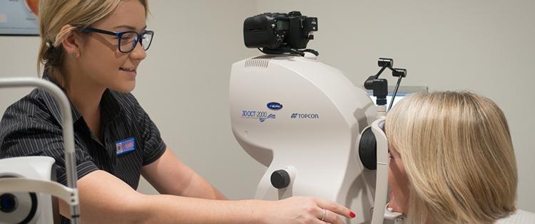
An OCT scan is a quick, painless and non-invasive imaging technique. The scan uses light to produce very high-resolution cross-sectional images of the layers of the retina. Images from some OCT machines will show blood vessels that are affected in AMD.
OCT scans are also used to monitor your individual response to any treatment you have.
Many optometrists now have OCT scanning technology.
Angiography
If your eye health professional suspects you have wet (neovascular) AMD, an ophthalmologist may perform a fluorescein angiogram.
Fluorescein is an orange dye. This dye is injected into the blood via a vein in your arm. The dye rapidly reaches the eye through the bloodstream and circulates through the retina. It will also highlight any abnormalities or damage to blood vessels.
A special camera will be used to take a series of images of your retina. This all takes only a few minutes.
In a very small number of cases, your ophthalmologist may perform an indocyanine green angiogram. This is where a green dye is injected into a vein in your arm in the same way as a fluorescein angiogram.


If you don’t already have an optometrist, our online search tool will help you find one in your area. You can even book your appointment online.
Find an optometrist
